Advanced Magnetic Resonance Imaging and Machine Learning Based Product Development for Noninvasive Detection of Genetic Subgroups of Brain Membrane Tumors
Advanced Magnetic Resonance Imaging and Machine Learning Based Product Development for Noninvasive Detection of Genetic Subgroups of Brain Membrane Tumors
Funded by TÜBİTAK 1001 Grants (119S520)
Principal Investigator: Esin Öztürk Işık
Co-Investigators: Alp Dinçer, Koray Özduman, Alpay Özcan, Ayça Ersen Danyeli, Murat Şakir Ekşi, Özge Can
Researchers: Abdullah Baş, Asım Samlı, Banu Saçlı Bilmez, Buse Buz, Esra Sümer, Gökçe Hale Hatay, Sena Azamat
Abstract
Brain membrane tumors, or meningiomas, are the most common brain tumors with a 35% incidence rate in adults. Meningiomas are more common in older people, and it is becoming a major health problem in our ever-aging society. While 80% of meningiomas are grade I and benign, grade III meningiomas tend to grow rapidly and spread to the surrounding tissue. In recent years, various genetic biomarkers for better understanding brain tumor biology have been identified, and they have been added to the classification criteria for brain tumors. These genetic biomarkers have become increasingly important in clinical practice today in diagnosing brain tumors, and to more accurately predict tumor biology, treatment responses, recurrence patterns, and survival rates. It is known that more than 60% of meningomas occur as a result of neurofibromatosis type 2 (NF2) gene changes. Molecular studies have shown that this molecular subgroup with NF2 mutations, which constitutes the majority of meningiomas, carry chromosomal instabilities, and they become malign, invade surrounding tissues, and turn into fast growing tumors. The knowledge of NF2 mutations in meningiomas prior to the application of treatment, such as surgery or radiotherapy, has the potential to significantly affect the treatment decision. However, these mutations are usually limited to the active tumor site, and could only be evaluated by pathological and molecular biological tests conducted on the tumor tissue after surgical removal. There is currently no product that might noninvasively provide NF2 mutation information in meningiomas. In recent years, advanced magnetic resonance imaging (MRI) based machine learning classification methods have gained great momentum in brain tumor studies. Diffusion tensor imaging (DTI), perfusion MRI, proton MR spectroscopic imaging (1H-MRSI), and susceptibility weighted imaging (SWI), provide information about white matter tract changes, perfusion and metabolic abnormalities, and magnetic susceptibility variations. Additionally, radiomic features of MRI modalities provide further hidden quantitative information about data characteristics. There has been a great interest in MRI-based classification of diseases using machine learning, and more recently deep learning methods. The aim of this study is to develop a machine learning based product, for detection of a radiological signature specific to the NF2 molecular subgroup of meningiomas, using advanced MR imaging and radiomic features. Preoperative diagnosis of NF2 molecular subtype of meningiomas by machine learning methods based on non-invasive MRI techniques will contribute to appropriate treatment planning and improvement of patient health.
Keywords: Brain membrane tumor, meningioma, magnetic resonance imaging, neurofibromatosis type 2 (NF2), machine learning, deep learning
Website of the tool: https://github.com/Computational-Imaging-LAB/IRIS-MRS-AI
Objectives:
- Develop a deep learning platform capable of classifying NF2 and S100-dependent subgroups of meningeal tumors using advanced MRI techniques, aiming for a minimum success rate of 80%.
- Investigate the effectiveness of individual advanced MRI methods in meningeal tumor classification using deep learning approaches and compare them with standard machine learning methods.
- Share the deep learning platform with the international scientific community through GitHub, expanding the scope of research in this field and contributing significantly to MRI-based understanding of the genetic variations of meningeal tumors.
- Enhance our country's knowledge and expertise in brain tumors, medical imaging, and deep learning by training specialized researchers in these areas.
Results:
- Magnetic Resonance Spectroscopy
- High grade meningiomas were found to have statistically significantly higher Glyc/tCr, Ins+Glyc/tCr and (Lip13a+Lip13b)/tCr values. Ins+Glyc/tCr value was more useful when used with (Lip13a+Lip13b)/tCr, (MM09+Lip09)/tCr, (MM14+Lip13a+Lip13b)/tCr to preoperatively determine the pathological grade using machine learning algorithms.
- Lower Ins/tCr and (Lip1a+Lip13b)/tCr were observed in hyperostotic meningiomas, and these metabolites were also found to be significant in detecting hyperostosis with machine learning algorithms in meningiomas. The ratios of Ins and (Ins+Glyc) to tCr were found to be lower in these tumors located at the base of the skull, which are called more benign.
- Our results showed that despite the small patient population, it is possible to detect NF2 loss in meningiomas with the help of single voxel proton MRS findings and machine learning or 1D-CNN model with 88.9% accuracy. Identification of NF2 loss using a deep learning model instead of classical machine learning algorithms increased the classification accuracy and reduced the computational load.
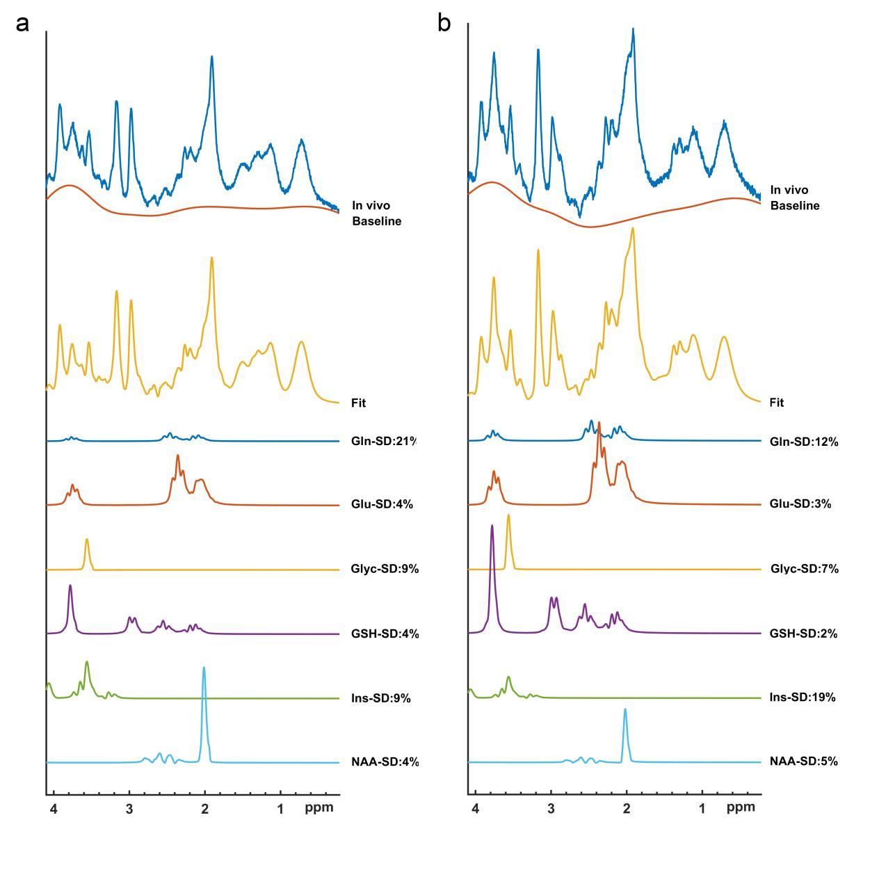
Figure. MR spectroscopic data of grade I (a) and grade II (b) meningioma patients with LCModel results.

Figure. Comparison of metabolite concentration values between meningiomas with and without NF2 loss (NF2-L). PCr + Cr = tCr, PCho + GPC = tCho, Glu + Gln = Glx, NAA+NAAG=tNAA.
- Susceptibility weighted imaging
- Meningiomas with S100 protein expression (S100+) tend to have a lower WHO grade than the S100- group, consistent with higher SWI values and more fibrous/transitive histopathological subtype findings.
- The heterogeneity of the signal intensity curve (standard deviation, entropy, and kurtosis) at the tumor site and the minimum signal intensity value reflecting the strength of the signal loss at the tumor site have been shown to correlate with progression-free survival time.
- The simultaneous evaluation of the SWI series with deep learning methods together with traditional MR methods (T1 weighted and FLAIR) was able to detect NF-2 gene loss with 76% accuracy.
- Radiomics
- It has been shown that the texture features are associated with the degree of meningioma.
- According to the results obtained from Friedman and Wilcoxon statistical tests in the evaluation of the consistency of radiomic shape features in T2 weighted images of meningiomas, significant differences were observed in most shape-based radiomic features. The ICC results showed the stability of all shape features except the sphericity index to the M1, M2, M3 preprocessing steps. This study demonstrated the high stability of the shape radiomic features in different smoothing filters and interpolation strategies.
- In this study, LASSO selected 6 important radiomic features while minimizing variation over 100 repetitions by generalizing the feature selection process.
- In the estimation of recurrence status in meningiomas with radiomics extracted from post contrast T1 weighted MRI data, skewness of both original and log filtered images, gray level association matrix (GLCM) cluster shadow, and minimal wavelet filtered image were found to be significantly associated with meningioma recurrence. Higher skewness values reflect more aggressive tumor biology and were significantly associated with recurrence, while GLCM cluster shadow and minimal post contrast T1 weighted images were associated with recurrence-free surveillance.

Figure. Distribution of predictive features (selection frequency >80%) selected in the two groups to distinguish between NF2-L and NF2-NL conditions. (*P<0.008)
- Diffusion Tensor Imaging
- Patients with S100 immunopositive had higher FA and RA ,and lower λ3 than patients who were not S100 immunopositive.
- Patients with loss of NF2 copy number had higher FA and RA, but lower λ2 when compared to those without.
- When deep learning models were used, ADC and λ3 maps obtained the highest accuracy in the S100 immunopositivity estimation in the test set, with 64.3% accuracy. In NF2 copy number loss, ADC and λ1 maps achieved 85.7% accuracy.
- Perfusion Weighted MRI
- Meningiomas with S100 protein expression resulted in lower rCBV values than meningiomas without S100 protein expressions.
- Meningiomas with the NF2 copy loss group resulted in lower rCBV values than NF2 no loss group.
- Deep Learning algorithms were evaluated.
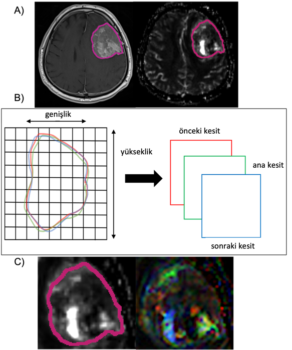
Figure. Deep learning application. A) Tumor mask in T1 post-contrast image and tumor mask superimposed on rCBV image, B) Method of transferring information from 3 slices to 3-channel image and C) 3-channel image generated from segmented tumor region.
- T2-weighted MRI
- The hybrid deep learning model achieved the best accuracy with 91% in the validation set and 83% in the test set (specificity=86%, sensitivity=78%) when majority voting was used as the accuracy criterion.
- Pretrained efficientnet-B2 network was chosen as the best performing architecture in the fine tuning procedure of the hyperparameter optimization.
- Generative Adversarial Networks
- In the study without GAN, 209 different slices were used randomly from patients (103 NF2-L, 106 NF2-NL) and 69.4% accuracy (64.1% sensitivity, 74.5% specificity) was obtained using post contrast T1-weighted images. To examine the effect of GAN, synthetic images were produced with GAN and 79.4% accuracy (75.7% sensitivity, 83% specificity) was reported with 209 randomly selected sections.
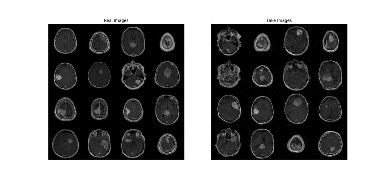
Figure. Real post contrast T1 slice images (left) and synthetic post contrast T1 slice images produced using GAN.
- Deep Learning Tool (IRIS-DL)
- Conventional and widely used model algorithms and auxiliary algorithms used in a typical machine learning flow have been added to the tool.
- Multi-layer perceptron (MLP), Convolutional Neural Networks (CNN), and Transformers will be added to make this work available to wider audiences and to better use the potential of artificial intelligence with IRIS-DL.
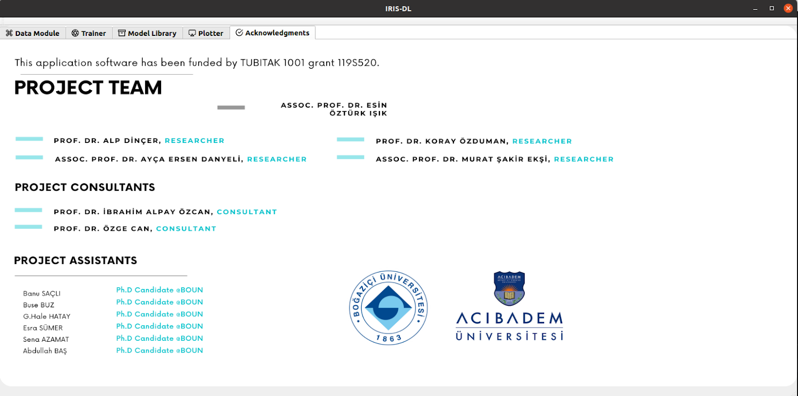
Figure. Screenshot of the IRIS-DL Team screen
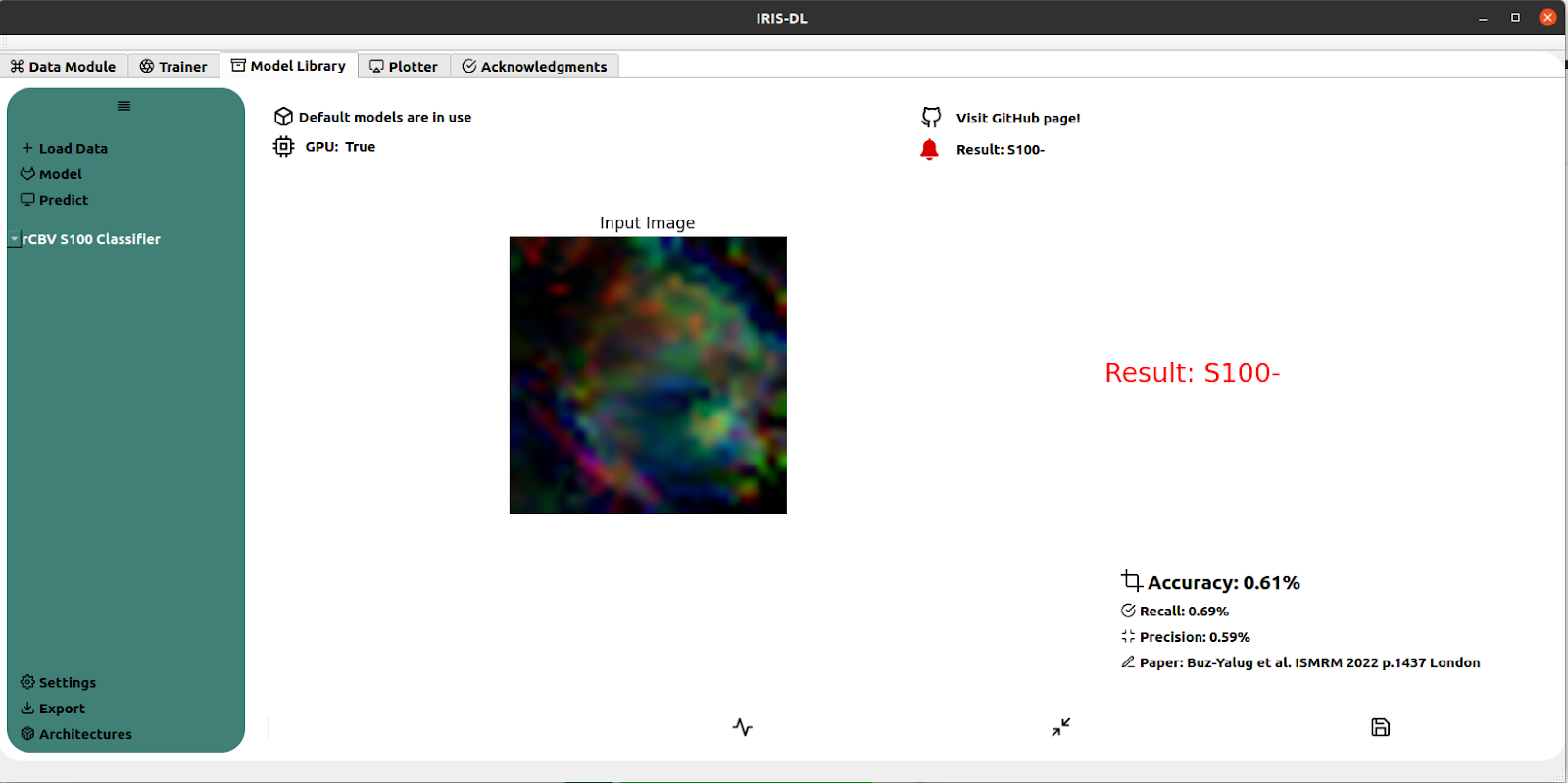
Figure. Detection of S100 expression with rCBV data and deep learning model.
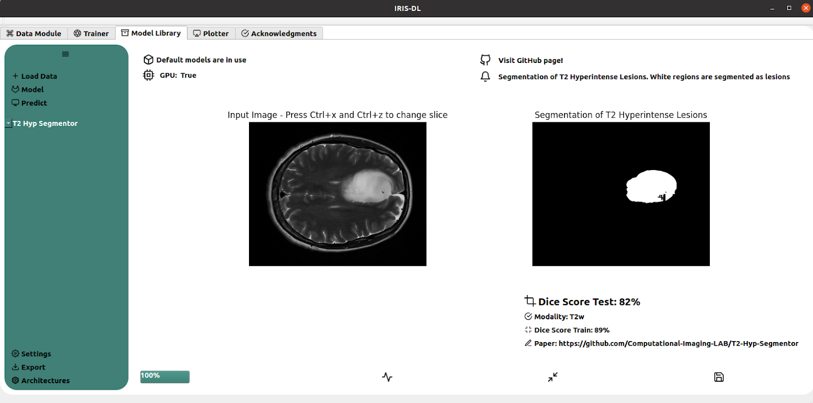
Figure. Tumor segmentation using 2DCNN-Unet2D over T2-weighted images.
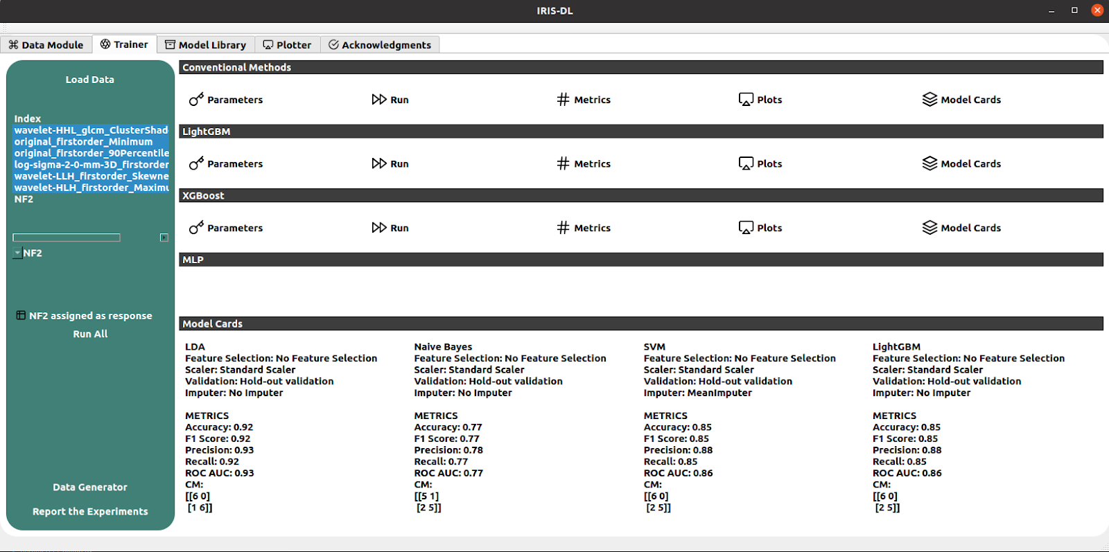
Figure. Model training interface
DISSEMINATION
Organized Workshops
‘Magnetic Resonance Spectroscopy Training’, organized with Gokce Hale Hatay, Banu Sacli Bilmez and Abdullah Bas. Bogazici University, March 19, 2022
Invited Speeches
- Ozturk-Isik E. Metabolites by MRS/I. ISMRM 2023. Educational session: ‘Quantifying Spins from Head to Toe’. June 3, 2023 (invited talk)
- Ozturk-Isik E. Imaging Physics - MRI of Gliomas. SPARK ACADEMY AFRICA-BRATS BRAINHACK 2023. May 24, 2023 (invited talk)
- Ozturk-Isik E. Detection of Genetic Mutations in Brain Tumors using Advanced Magnetic Resonance Imaging-based Machine Learning. Turkish Health Institutes (TÜSEB), Turkish Cancer Institute (TKE), Innovative Diagnostic Methods in Cancer Seminar. April 6, 2023. (invited talk)
- Ozturk-Isik E. The Role of Machine Learning in Brain Tumor Classification and Detection of Genetic Mutations. 18th Congress of Neurosurgery. Antalya, Turkey, October 27, 2022 (invited talk)
- Ozturk-Isik E. Pearls of Dublin I: Genomics – Integrating Genomics into Neuro-oncology. GliMR 2.0 3rd Annual Meeting. Kuşadası, Izmir, Turkey, September 28, 2022 (invited talk)
- Ozturk-Isik E. Genomics – Integrating Genomics into Neuro-oncology. GliMR Training School – Artificial Intelligence in Neuro-Oncology. Dublin, Ireland, July 25, 2022 (invited talk)
- Sümer E., Radiogenomics applied (Hands-on)–Pyradiomics, GliMR Training School, July 25 2022, Dublin, Ireland (invited talk)
- Ozturk-Isik E. How do Radiologists and Engineers Collaborate? An Engineer's Perspective. TMRD 2022. May 26, 2022. (invited talk)
- Ozturk-Isik E. How Can AI Help for MRS? ISMRM 2022. Educational session: ‘Steady State MRS’. London, UK, May 8, 2022 (invited talk)
- Ozturk-Isik E. Innovative Approaches in Medical Imaging. March 30, 2022. Isik University, Biomed IV. (invited talk)
- Ozturk-Isik E. Machine Learning on MRI Images for the Detection of Genetic Mutations in Brain Tumors. TÜSEB Artificial Intelligence in Healthcare Mini Symposium Series – 6. December 29, 2021 (invited talk)
- Ozturk-Isik E. Prediction of Recurrence Risk of Meningiomas with MRI. TURNOG. December 12, 2021 (invited talk)
- Ozturk-Isik E. Machine Learning-Based Classification of Genetic Mutations in Brain Tumors for Determining Prognosis: MR Spectroscopic Imaging Analysis. Acıbadem Üniversitesi Nörolojik Bilimler Toplantısı. January 25, 2020. (invited talk)
Conference Proceedings
International Proceedings:
- Azamat S, Buz-Yalug B, Ozcan A, Ersen Danyeli A, Pamir MN, Dinçer A, Ozduman K, Ozturk-Isik E. A Pretrained CNN Model Using Multiparametric MRI to Identify WHO Tumor Grade of Meningiomas. International Society for Magnetic Resonance in Medicine. International Society for Magnetic Resonance in Medicine. Toronto, Canada, June 3-8, 2023 (digital poster)
- Sümer E, Türe OA, Şengöz M, Pamir MN, Dinçer A, Özduman K, Ozturk-Isik E. Correlation of MRI Radiomics Features and Recurrence After Gamma Knife Treatment in Parasagittal and Falx Meningiomas. International Society for Magnetic Resonance in Medicine. Toronto, Canada, June 3-8, 2023 (digital poster)
- Dindar SS, Buz-Yalug B, Tan K, Danyeli AE, Can O, Pamir N, Dincer A, Ozduman K, Kahya YP, Ozturk-Isik E. Prediction of NF2 Loss in Meningiomas Using Synthetic T1-Weighted Contrast Enhanced MRI Generated by Deep Convolutional Generative Adversarial Networks. International Society for Magnetic Resonance in Medicine. Toronto, Canada, June 3-8, 2023 (digital poster).
-
Bas A, Danyeli AE, Can O, Ozduman K, Dincer A, Ozturk-Isik E. Identification of NF2 loss in meningiomas using T2-weighted MRI and Deep Learning. International Society for Magnetic Resonance in Medicine. Toronto, Canada, June 3-8, 2023 (digital poster)
-
Bas A, Sacli-Bilmez B, Buz-Yalug B, Sumer E, Azamat S, Hatay GH, Danyeli AE, Can O, Ozduman K, Dincer A, and Ozturk-Isik E. IRIS-DL: A Deep Learning Software Tool for Identifying Genetic Mutations in Gliomas and Meningiomas. International Society for Magnetic Resonance in Medicine. Toronto, Canada, June 3-8, 2023 (oral presentation)
-
Sümer E, Ozturk-Isik E, A radiomics pipeline for neuro-oncological research. GliMR 3rd Annual Meeting. Kuşadası, Turkey, September 28-30, 2022 (oral presentation)
-
Buz-Yalug B, Ersen Danyeli A, Ekşi MŞ, Tan K, Can Ö, Yakicier C, Pamir MN, Dincer A, Ozduman K, Ozturk-Isik E. Differentiation of NF2 loss and S100 presence in Meningioma using Dynamic Susceptibility Contrast MRI with Machine Learning Approach. International Society for Magnetic Resonance in Medicine. London, UK, May 7-12, 2022 (digital poster)
-
Azamat S, Buz-Yalug B, Baş A, Ozcan A, Ersen Danyeli A, Pamir MN, Dinçer A, Ozduman K, Ozturk-Isik E. Susceptibility Weighted MRI for Predicting Critical Developmental Regulatory S100 Proteins in Meningiomas at 3T. International Society for Magnetic Resonance in Medicine. London, England, UK. May 07-12, 2022. (digital poster)
-
Sacli-Bilmez B, Bas A, Tan K, Ersen Danyeli A, Can Ö, Yakicier C, Pamir MN, Dincer A, Ozduman K, Ozturk-Isik E. Identification of NF2 loss in meningiomas using 1H-MRS at 3T. International Society for Magnetic Resonance in Medicine and Biology. London, UK, May 7-12, 2022 (digital poster)
-
Sumer Esra, Tan K, Ersen Danyeli A, Can Ö, Yakicier C, Pamir MN, Dincer A, Ozduman K, Ozturk-Isik E. Meningiomas with NF2-Loss Exhibit Strong Radiomics Correlations on Contrast Enhanced T1-Weighted MRI at 3T. International Society for Magnetic Resonance in Medicine and Biology. London, UK, May 7-12, 2022 (online oral power pitch presentation)
-
Bas A, Tan K, Ersen Danyeli A, Can Ö, Yakicier C, Pamir MN, Dincer A, Ozduman K, Ozturk-Isik E. Identification of S100 Immunopositivity on T2-weighted MRI Using Deep Learning. International Society for Magnetic Resonance in Medicine and Biology. London, UK, May 7-12, 2022 (digital poster)
-
Sümer E, Arpak A, Pamir MN, Dinçer A, Özduman K, Ozturk-Isik E, Assessment of Stability of Radiomic Shape Features on T2-Weighted Images of Meningiomas, The European Society for Magnetic Resonance in Medicine and Biology. Virtual Meeting, Oct 7-9, 2021, p.182. (digital poster)
-
Sümer E, Pamir MN, Dinçer A, Özduman K, Ozturk-Isik E, Classification of Low- and High-Grade Meningiomas Using Radiomics Features of Post-Contrast T1-Weighted MRI, The European Society for Magnetic Resonance in Medicine and Biology. Virtual Meeting, Oct 7-9, 2021, p.181. (digital poster)
-
Halilibrahimoglu H, Buz Yalug B, Kaykayoglu A, Ersen Danyeli A, Eksi M.S, Yakicier C, Pamir MN, Dincer A, Ozduman K, Ozcan A, Ozturk–Isik E, Classification of High- and Low-Grade Meningiomas Using Diffusion Anisotropy Indices with Deep Learning. The European Society for Magnetic Resonance in Medicine and Biology. Virtual Meeting, Oct 7-9, 2021, p.184-185. (digital poster)
-
Sacli-Bilmez B, Bas A, Ersen Danyeli A, Ekşi MŞ, Tan K, Can Ö, Yakicier C, Pamir MN, Dincer A, Ozduman K, Ozturk-Isik E. 1D-CNN for grading of meningiomas using Proton Magnetic Resonance Spectroscopy. The European Society for Magnetic Resonance in Medicine and Biology. Virtual Meeting, Oct 7-9, 2021, p.178-179. (digital poster)
-
Sacli-Bilmez B, Ersen Danyeli A, Ekşi MŞ, Tan K, Can Ö, Yakicier C, Pamir MN, Dincer A, Ozduman K, Ozturk-Isik E. Correlations of Single Voxel 1H-MRS Findings with Tumor Biology in Meningiomas. International Society for Magnetic Resonance in Medicine. Virtual Meeting, May 15-20, 2021, p.954. (digital poster)
-
Sümer E, Tek E, Pamir MN, Şengöz M, Dinçer A, Özcan A, Özduman K, Ozturk-Isik E. Association of Radiomics Based Tumor Shape Irregularity Measures and Gamma Knife Dose Planning Indices in Vestibular Schwannomas. Society of Neuro-Oncology (SNO) 25th Annual Meeting. Austin, Texas, USA, November 19-22, 2020, p.NIMG-57. (digital poster)
National Proceedings
- Sümer E, Pamir MN, Dinçer A, Özduman K, Ozturk-Isik E, Classification of Low- and High-Grade Meningiomas by Radiomic Characteristics Calculated from Contrast-Enhanced T1-Weighted MR Images, TMRD 2022, Ankara, Turkey, March 26-27, 2022. (oral presentation)